Article Text

Abstract
There are limited curricular options for medical students to engage in art-making during their training. Yet, it is known that art-making confers a variety of benefits related to learning. This qualitative study utilises a visual methodology to explore students’ art-making in the context of the cardiovascular sciences. The existence of a multiyear repository of medical/dental student generated, cardiac-inspired art, collected over 6 years, provided the opportunity to explore the nature of the art made. The aim was to categorise the art produced, as well as the depth and breadth of understanding required to produce the art. The data set included a wide variety of titled art (paintings, photographs, sketches, sculptures, collages, poetry and music/dance). Systematic curation of the collection, across all media, yielded three main categories: anatomical renderings, physiology/pathophysiology renderings and kinesthetic creations (music/dance/tactile). Overall (medical and dental) student-generated art suggested a high level of content/process understanding, as illustrated by attention to scientific detail, integration of form and function as well as the sophisticated use of visual metaphor and word play. Dental students preferentially expressed their understanding of anatomy and physiology kinesthetically, creating art that required manual dexterity as well as through choreography and dance. Combining art-making with basic science curricular learning invited the medical and dentistry students to link their understanding to different modes of expression and a non-biomedical way of knowing. Subsequent incorporation of the student-generated cardiac art into lectures exposed the entire class to creative pictorial expressions of anatomy, physiology and pathophysiology.
- art and medicine
- cardiology
- medical education
- music
- dance
Statistics from Altmetric.com
Background
Medical students have a wide variety of academic and non-academic talents that were fostered during the years prior to entering medical school. It is no longer unusual for students accepted to North American medical schools to have completed an undergraduate degree in arts or music, rather than science. Despite the fact that medical and dental students bring an interest in arts and humanities into their professional training, there are few opportunities for acting on these interests from within their programmes. Yet benefits of medical student art-making have included decreased stress, a better work–life balance and exploration of personal and professional identity.1–3 Creative art was shown to promote a reflective, humanistic practice.4 In addition, art-making (as distinct from art-observation) by medical students has the potential to enhance interest in, solidify and integrate, learning.1 5–7 Cardiac art made by medical students in Nepal demonstrated a strong conceptual understanding of cardiac sciences.8 Artist statements that accompanied submissions to a national medical education art exhibition described why art-making was important to them. A qualitative analysis of the statements identified eight themes, among them attributes of learning (such as comprehensiveness, retention and enjoyment) that are enhanced through artistic expression.5
Art-making within a science context may therefore offer medical students an alternative, non-biomedical approach to learning about physiology and anatomy. Art-making allows learners to access tacit knowledge—defined by Polyani as knowledge that exists beyond conscious awareness—and to express more than they could by using words alone.9 10 Shklovsky describes the major function of literature and art in general is to disrupt automatic recognition and to allow the learner to look at the familiar in new ways.11 Embedding education (medical or otherwise) with art-making promotes self-exploration and integration with self and others and their place in the outside world.9 12
It is important to note that art-making for students does not take place in a vacuum, rather new ways of knowing depend at least in part on the background of the learner. Students enter medical school with an established world view, personal identity and a personal aesthetic established during and prior to their premedical school education. In art-making, there is a natural back and forth between prior knowing and new situations that influences and transforms learning.13 Art-making in the context of learning medicine therefore provides students with the opportunity to reconnect and integrate their learning with their past creative selves.4
As the director of cardiovascular learning during the first-year curriculum in a medical school, I provided medical and dentistry students (whose first year curricula ran concurrently) with the opportunity to express artistically their learning of, and appreciation for, the cardiovascular system. This online multiyear repository of cardiac-inspired art is called ‘The Heartfelt Images’ (found in the Art Gallery at www.teachingmedicine.com). The call for original art was broad and asked only that the art represent the cardiovascular sciences.
The present study was designed to take advantage of the presence of this multiyear repository and examine it for evidence of students engaging in a different way of learning and/or knowing within the confines of a single discipline (cardiovascular sciences). The aim of this exploration was to curate the collection, in an effort to reveal the nature and level of scientific understanding required by the students in order to create their art.
The data
Images examined in the present study from the Heartfelt Images repository were restricted to the last 6 years of submissions. This was done since students whose art was created after 2011, were either still in the medical/dentistry programme or only recently graduated, and thus were easier to contact for the purpose of obtaining consent (University Ethics Board). All images produced during the time period (between 2011 and 2016) were included in the analysis. A total of 262 students submitted 345 pieces of art to the ‘Heartfelt Images’ repository during that time period. All of the students who submitted art (241 medical students, 21 dentistry students) were enrolled as full-time, first-year medical or dentistry students in our programme at the time of submission.
The data set for this analysis included the images and their titles. A subset (the most recent years) of images were submitted with artist statements. While the artist statements did not contribute to the curatorial analysis, some details from the artist’s statements were added to the manuscript (eg, Spirit of the Heart and Take a Bite out of Heart Disease) for the purpose of explaining their cardiovascular significance. Eight images were highlighted in the manuscript along with the artists’ names, and written consent to include the art and their names was obtained from the artists.
Participation in the art-making was entirely voluntary and not linked to curricular assessment. The submission rate represented 16.7% and 7.4% of the total medical and dentistry classes, respectively. Submission numbers from males and females were similar.
For both medical and dentistry students, the majority of the cardiac art was submitted in the form of photographs. Second to photographs were sculptures/installations (more so by dentistry students), and thirdly, sketches. Audio-visual submissions, paintings, collages and poetry made up the remaining submissions.
The analysis
To curate a collection of art is to present the items using one’s professional or expert knowledge. This curatorial analysis was conducted through the author’s dual lens of multidecade teaching expertise in the cardiac sciences and amateur photographic practice. The methodology used in this study was exploratory in nature, rather than a determined qualitative analysis.8 That said, Rose’s criteria of ‘close looking’ and reflexivity were adhered to in this study. Two criteria cited by Rose for a critical approach to visual methodology include: taking images seriously—through close looking, considering researchers own way of looking at images (reflexivity).14
The curatorial aim was to reveal the nature of student understanding of the cardiovascular science necessary for creation of the art. Curation of the collection began with a close examination of each piece of art and assignment of broad scientific descriptions (eg, anatomy, physiology or pathophysiology). Second, specific identifiers (eg, literal, imaginative, metaphorical, humorous, linked to nature, linked to health, kinesthetic and so on) emerged organically. This process was repeated in an iterative fashion until the list of identifiers was exhausted. As a result of steps 1 and 2 in the curation, three main categorisations emerged:
anatomical renderings
physiology/pathophysiology renderings
kinesthetic creations (music/dance/tactile).
Categorisation occurred across all media. These categories were not mutually exclusive. Some pieces of art fit into more than one category.
Main catagories
Anatomical renderings
This was a main category for both medical and dentistry students. Images of cardiac anatomy in this category included found items; that is, students saw objects or scenes in the urban or natural world that were either quite faithful to their understanding of cardiac anatomy, or in many cases represented more ‘stylized’ valentine hearts. In addition, and different from found items, students actively assembled a variety of materials into anatomically accurate representations of hearts. Finally, many students represented cardiac anatomy imaginatively or metaphorically.
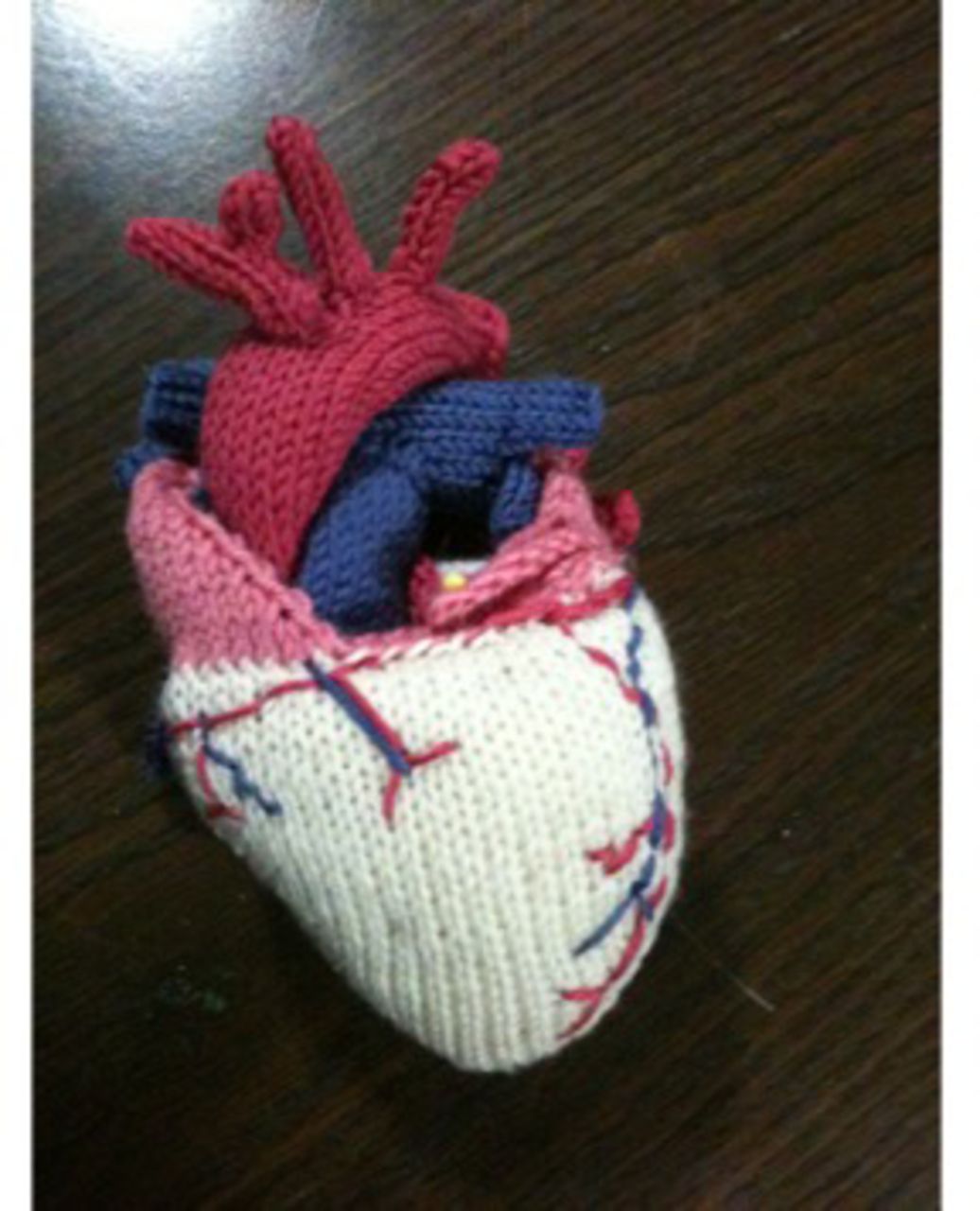
An example of the many found items from nature that were submitted was one by D. Barnes (figure 1). Barnes photographed patterns in the sand made as waves drained from a beach that for him represented histological striations in the heart wall known as Trabeculae Carnae. Representing art that was created (rather than found) was a hand knitted, anatomically accurate, three-dimensional heart made out of wool by A. Miners (figure 2) entitled ‘Softhearted’. Students alternatively used clay, wire and various kinds of food to construct cardiovascular anatomy. C. Wong (figure 3) artfully used his body, in a classic yoga pose, to produce a stylised heart shape and called it Namast’ay Healthy, thus promoting heart-healthy practices. At the imaginative end of the spectrum, students took artistic and creative licence to depict cardiovascular anatomy metaphorically. A metaphor, beyond being a figure of speech, can be an object that is used, or regarded as being used, to represent something else.15 Thus, A. Miners ‘Softhearted’ mentioned above, incorporated metaphorical content as indicated by the work’s title, while M. Maruyama (figure 4) imagined four animals (whale, seal, fox and bird) combining to form an anatomically accurate heart in a painting she titled ‘Spirit of the Heart’. The two water-dwelling animals (whale and seal) represented the deoxygenated blood that flows through the right side of the heart, while the two land creatures (fox and bird) represented the oxygenated blood flowing through the left side of the heart. This painting was an homage to Maruyama’s Irish/Japanese heritage as well as to the First Nations Aboriginal culture in British Columbia. While encased in artistic design, the attention to cardiovascular detail in Maruyama’s painting was evident. All the anatomical features were present, and yet seamlessly incorporated into the design of the animals (eg, whale fins and fox ears as heart valve leaflets, bird feathers as carotid arteries, whale and fox teeth as electrical conduction fibres).

D. Barnes’s photograph entitled ‘Trabecula Carnae’ illustrating cardiac wall histological striations as the sand patterns made as waves drained from a beach.

A. Miner’s knitted wool sculpture entitled ‘Soft-hearted’ of an anatomically accurate, three-dimensional heart.

C. Wong artfully used his body in a classic yoga pose to produce a heart shape and called it ‘Namast’ay Healthy’.

M. Maruyama’s painting entitled ‘Spirit of the Heart’ depicts an anatomically accurate heart as four entwined spirit animals (whale, fox, seal and bird).
Physiology and pathophysiology renderings
This was another main category for both medical and dentistry students. Students artistically and creatively translated complex physiological and pathophysiological phenomena into photos, sketches, paintings and installations.
Students again used their ‘cardiovascular lens’ to look at nature and the urban landscape; however, the art represented a physiological/pathophysiological process, rather than static anatomy. One example was a ‘Re-entry Circuit’, which is a circular electrical pathology in the heart that results in a rapid and irregular heart rhythm. M. Benusic (figure 5) used time-lapse photography to capture the lights of cars as they entered and exited a traffic roundabout in order to artistically conceptualise an urban re-entry circuit.
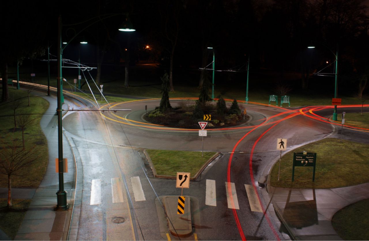
M. Benusic used time-lapse photography to capture the lights of cars as they entered and exited a traffic roundabout in order to artistically conceptualise an urban ‘Re-entry Circuit’.
As separate from seeing physiology/pathophysiology in the urban or natural landscape, students also represented the physiological function of the heart metaphorically. I. Jokic (figure 6) represented the very slow beating of the heart (termed bradycardia) as a snail moving ponderously across the landscape, while the mountain peaks on the horizon represented the infrequent QRS complexes seen in the ECG of a patient with bradycardia.
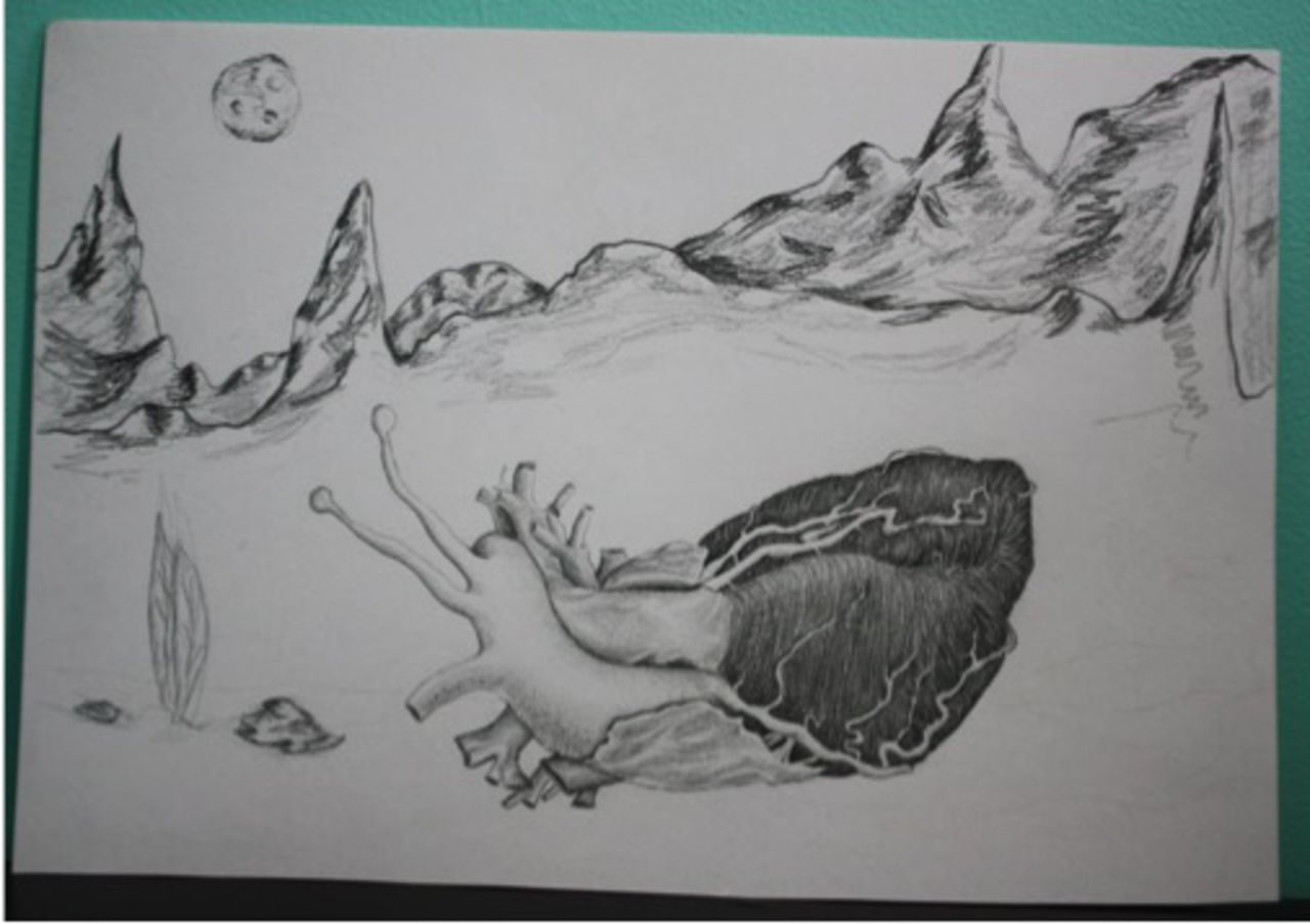
I. Jokic’s sketch entitled ‘Bradycardia’ represented the very slow beating of the heart as a snail.
Homonyms (words with dual meanings) in some of the titles shed light on the metaphorical nature of a number of physiological and pathophysiological renderings. D. Wan’s (figure 7) piece entitled ‘Arrested’ illustrated several different levels of understanding. It showed the blood supply to the heart that, when compromised, could produce a cardiac emergency known as ‘Cardiac Arrest’. That image was combined with a drawing of a closed fist, often used to illustrate the anatomical size of a person’s heart. In addition, a closed fist is the gesture patients often use when describing the squeezing feeling they may have leading to cardiac arrest. To complete the image, Wan included a wrist wearing traditional police handcuffs.

D. Wan combined a sketch of the blood supply of the heart with a drawing of a clenched fist and a wrist encircled with handcuffs. Taken together, the image was entitled ‘Arrested’.
In effect, all these medical/dental student artists whether focusing on static anatomy, or physiological and pathophysiological processes, were looking at the familiar with new eyes, something Shklovsky referred to as ‘estrangement’.11 Seeing the world through a ‘cardiovascular lens’, and thinking metaphorically may ‘educate attention’ thus allowing for deeper seeing or appreciation.16
Duit posited in ‘On the Role of Analogies and Metaphors in Learning Science’ that the element of surprise or anomaly (eg, the heart as a snail shown in the present study), make metaphors significant in the learning process and that their ‘generative power makes them potentially valuable tools in conceptual change learning’.17 Llewellyn et al regarded metaphors as a form of creativity that ‘far from obscuring reality and knowledge is a pathway to new understandings and potential solutions’.18 Clinical medicine is no stranger to metaphors as evidenced by their use in specialties like pathology and dermatology (eg, ‘pea soup diarrhea’ or ‘oat cell carcinoma’). When medical students move from text to the creation of a visual metaphor during the art-making process, they open themselves to the possibility of generative learning and may internalise a visual image that is more easily retrieved later.19 20
Students whose art fell in the category ‘Physiology/Pathophysiology Renderings’ seemed to be providing visual explanations for difficult physiological and pathophysiological concepts. This may have benefits for their learning. Bobek and Tversky compared student learning after creating visual explanations of scientific phenomena and concluded that ‘visual explanations map thought more directly than words and provide checks for completeness and coherence as well as a platform for inference, notably from structure to process’.21
Kinesthetic creations
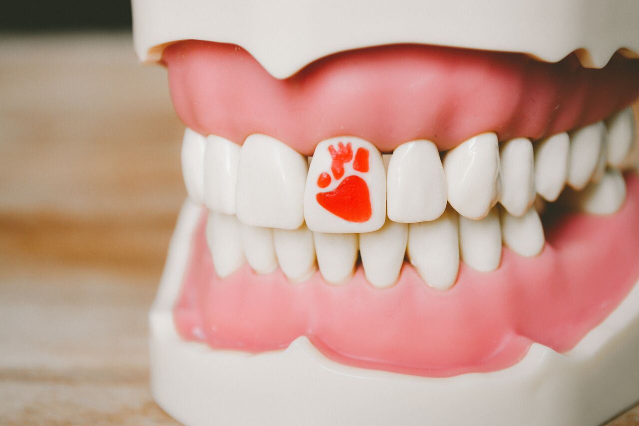
Students whose art was included in this category (largely dentistry students) illustrated a preference for creating art that involved a certain amount of manual dexterity, hand–eye coordination and movement. Some of the dentistry students used ‘typodonts’; these are educational models that allow students to ‘drill and fill’ teeth in order to practice their clinical skills. S. Bortolussi (figure 8) used a dental handpiece to engrave an anatomical heart into a tooth in a typodont and then back-filled it with red wax and called her submission ‘Take a bite out of heart disease’. The message here was the link between poor dental hygiene and the danger of heart disease. Using their art to advocate for better health extends the student’s own personal learning into a larger awareness of the human experience and an understanding of their responsibility as future practitioners.9

Bortolussi used a dental handpiece to engrave an anatomical heart into a tooth in a typodont and then back-filled it with red wax and called her submission ‘Take a bite out of heart disease’.
Dentistry students also employed music and movement to ‘perform’ cardiovascular concepts. A. Sotoodeh choreographed and recorded a performance of an original group salsa dance in order to illustrate normal cardiac rhythm. C. Chu composed and recorded an original score for Cello entitled ‘In a heartbeat’ that drew from a strong percussion beat in the background.
Students studying dentistry, like any collection of learners, have a variety of learning styles (visual, auditory, read/write and kinesthetic) and show a preference for multimodal learning.22 That dental students would be inclined towards tactile submissions should not be surprising given that one aspect of dental school selection includes their scores on the Dental Aptitude Test (DAT). As stated on the Canadian Dental Association website, ‘The DAT tests not only academic ability, but importantly, two and, three-dimensional visual perception and manual dexterity’.23
Role for art-making/art-viewing in class engagement
While it may seem like a limitation to engage only 16.7% and 7.4% of the total medical and dental class (respectively) in art-making, it could be problematic to make this compulsory. Bristol University is one of the few medical schools that embeds what they refer to as ‘Compulsory Creativity’ into their curriculum.24 The challenge inherent in such a programme, and what may deter other schools from requiring art-making within their curriculum, is the necessity for faculty to assess the creative work in a way that is transparent and defensible.25 The low participation rate in the art-making notwithstanding that does not mean that the entire class did not derive benefit. All students in the class were exposed annually to cardiac art made by their peers in an end of block art presentation. In addition, throughout lectures during the annual 5 weeks of learning about the cardiovascular system, student-generated cardiac art (from previous years) was shown to illustrate anatomical, physiological and pathophysiological concepts. These artistic images, when paired with scientific explanations, were meant to stimulate the students’ imagination and provide them with alternative (visual) ways of thinking about anatomy or physiology/pathophysiology. For the medical student artists, this showing of their pieces during lectures provided a ‘site of audiencing’ that allowed their peers to make their own interpretations of the images.14 This ‘picture is worth a thousand words’ strategy may find power in the picture superiority effect (ie, pictures generally show superior recognition relative to their verbal labels).26 The use of pictorial mnemonomy (interpretational pictures used to clarify difficult scientific concepts) was found to facilitate performance by students on reconstruction and application of information.27 No one has studied the impact on medical students of learning from text or text plus pictures; however, studies have reported the benefit of pictures added to text in patient literature in enhancing patients’ attention, comprehension, recall and adherence.28
Summary
This study describes a curation of cardiac-inspired art created by medical and dental students and contained in a multiyear, online repository. The aim of the curation was to reveal the levels of scientific understanding required by the students in order to create their art. Art generated in all categories suggested a high level of content/process understanding as illustrated by attention to detail, integration of form and function as well as the sophisticated use of visual metaphor and word play.
Through their art, medical and dentistry students saw cardiovascular anatomy and physiological and pathophysiological concepts in natural and urban landscapes. In effect, they looked at the world through a cardiovascular lens. Combining artistic expression with basic science curricular learning opened the door to involving other senses and invited the medical and dentistry students to link their understanding to different modes of expression. Subsequently, incorporating the student-generated cardiac art into lectures exposed the entire class to creative, pictorial expressions of anatomy, physiology and pathophysiology.
Future directions
Heartfelt Images, the annual call for cardiac art, is an ongoing endeavour that could prompt future research questions. What is the effect on learning of making versus capturing (as a photograph) the cardiac art? What is the effect of seeing the student generated art for the students in the class that did not participate in art-making? Are there barriers for students who do not see themselves as artists in participating in art-making, and if so, how might they be circumvented (or should they)? Lastly, it would be interesting to determine a mechanism for participation in art-making (in the context of disciplinary learning) as a curricular endeavour, thus necessitating an exploration of strategies for assessment.
Acknowledgments
I thank Dr Susan Cox (UBC) for insightful edits and comments in the final stages of manuscript preparation and Dr. J. Waechter for hosting the art on teachingmedicine.com. I am grateful to all the medical and dental students who submitted cardiac art to ‘Heartfelt Images’. Your creativity inspires me. In particular my thanks to the students whose art was highlighted in this article: medical students: D Barnes, A Miners, I Jokic, M Maruyama, M Benusic, D Wan and C Wong; dentistry student: S Bortolussi, A Sotoodeh and C Chu.
References
Footnotes
Twitter Follow Carol Ann Courneya @cacourneya
Contributors CAC planned and implemented the study and wrote the manuscript.
Competing interests None declared.
Ethics approval University of British Columbia Research Ethics Board.
Provenance and peer review Not commissioned; externally peer reviewed.